
")
Förster Resonant Energy Transfer
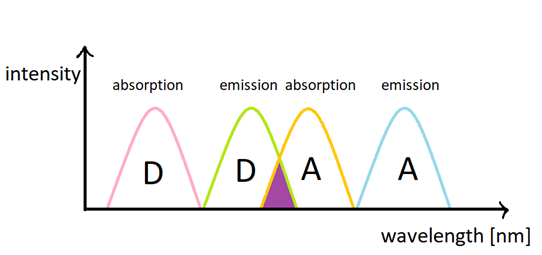
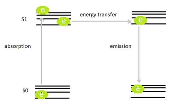
Förster Resonant Energy Transfer (FRET) is a generic method to study interaction between two fluorescent maieties - the donor (D) and the acceptor (A). Energy absorbed by the donor may be transfered non-radiatively to the acceptor due to dipol-dipol interactions under three circumstances :
(1) D emission overlaps spectrally with A absorption
(2) D and A stay in close proximity (<10 nm)
(3) D and A are properly oriented in space.
This 'spectroscopic ruler' phenomenon creates the basis for design of numerous biosensors to study e.g. conformation of proten, enzyme activity, DNA hybridization, antibody-antigen reactions. This methods may be used either for medical diagnostics or in fundamental studies to understand the processes occuring in living cells.
Whereas organic fluorophores (dyes, fluorescent proteins etc.) are commonly used for FRET sensing (as D or A), such lables display numerous drawbacks, such as (1) photobleaching, of D and A, (2) spectral bleed through between D/A excitation and emission channels, (3) low signal-to-background signal (D are excited with wavelengths suitable to photoexcited autofluorescence of biomolecules, (4) very short fluorescence lfietimes. LnNP and UCNP circumvent many of these drawbacks, but set new class of challanges, such as many-D-many-A detection, long riseitmes of D photoluminescence, recharging of D species throught upconversion process. We aim to study, understand and apply such new UCNPs based bio-detection materials and methods.
FRET effiency can be estimated following the equation below:
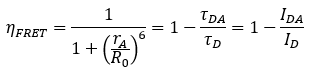
The ΙD and ΙDA are the integrated luminescence intensity of the donor without and with the presence of acceptor molecules. Similarly τD and τDA are luminescence lifetime of donor alone and of the donor accompanied by the acceptor.
Due to inverse 6th power distance dependence, the increased donor to acceptor distances can result in reduced FRET efficiency. To quantify this value experimentally one can measure either luminescence lifetime, donor luminescence intensity drop and acceptor fluorescence (if the acceptor is fluorescent).
We are interested in using lanthanide doped nanoparticles, and especially upconverting nanoparticles (UCNPs) as energy donors in RET based sensors. They have a few tens of nanometers in diameter. They demonstrated an exceptional capability to generate visible or far-red luminescence emission under NIR photoexcitation providing them unique advantages as FRET donors. The example of these compounds is NaYF4: Yb3+, Er3+. The first advantage of them is their high photostability. It is caused by ions Ln3+ because of the fact that 4f orbital electrons are shielded against the influence of the chemical environment by filled 5s and 5p orbitals. The next benefits are narrow emission bands, large anti-Stokes shifts and long decay times of luminescence. The long lifetime of the luminescence is due to the Laporte’s rule forbidden of the transition inside the f-shell. UCNPs are able to emit ultraviolet, visible or near-infrared light after absorbing NIR light.
We aim to study, understand and apply such new UCNPs based bio-detection materials and methods.
We design and the assess the virtual nanocrystal NP (VNP) models which lead to the new designs of UCNPs. Core-shell nanoparticles have an inner core structure and an outer shell made of different components. These particles have been of interest as they can exhibit unique properties. This nanoparticles are experimentally evaluated as donor NPs and compared to the simulations. Our studies help to understand the role of energy-transfer and energy migration between lanthanide ion dopants and how the architecture of core-shell UCNPs affects their performance as FRET donors to organic acceptor dyes. The structure and energy diagram of synthesized core-shell NPs we can see in the picture below (Fig.3).
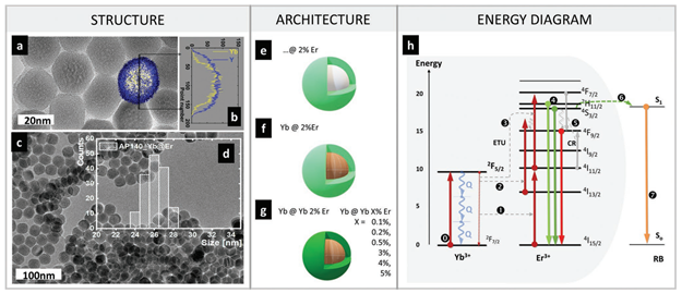
Fig. 3. The material properties (structure), synthesized core-shell NPs (architecture) and Yb3+-Er3+ upconversion based RET (energy diagram). Representative a) TEM, b) TEM-EDS, c) large field-of-view TEM, d) size distribution of Yb@Er Sample. The schematic description of the synthesized NPs: e) undoped-core-active-shell (…@2Er), f) sensitized-core-active-shell (20Yb@2Er), g) sensitized-core-active-shell (20Yb@20Yb2Er, and additional 0.1, 0.2, 0.5, 3, 4, and 5%); h) energy diagram of energy transfer Yb-Er3+ up-conversion based Er-to-RB RET.
We found interesting relationships, which confirm that specially designed core-shell architectures of UCNPs (with donor ions in the shell) are beneficial for UC-LRET sensitivity over conventional UCNPs (where donor ions are evenly distributed in the whole volume). Moreover, we demonstrated that the different photoexcitation scheme, may have their parasitic role in decay based UC-LRET sensing.
Moreover we studied Förster resonance energy transfer between nanoparticles of up-conversion lanthanide crystals as donors and quantum dots as acceptors. We have demonstrated that this system possesses the essential features required for FRET applications. These include high D-A spectral overlap and satisfactory quantum yield of the donor with no acceptor present. Both acceptor emission and donor luminescence lifetime decrease were experimentally demonstrated and were in good agreement with the theoretical Förster distance value.
In our work focused on the effect of Tm3+ ion concentration on FRET efficiency, we compared two classical approaches for evaluating FRET: donor ion emission quenching and shortening of the excited state lifetimes of donor ions in the presence of an acceptor. By directly conjugating dyes to the surface of nanocrystals, we achieved high energy transfer efficiencies of approximately 90% and 40%, respectively, for the ATTO 488 dye. However, these approaches yield different numerical values due to the complex nature of upconversion processes, energy migration within the Yb3+/Tm3+ lattice, and repopulation of the excited levels of Tm3+ ions, and therefore cannot be interpreted independently. We demonstrated that the most sensitive measure of FRET was ratiometric detection, based on the ratio of acceptor emission intensity to donor emission intensity (in the presence of the acceptor) within strictly defined spectral windows (500–614 nm and 435–485 nm). Although this method does not provide a detailed description of the FRET mechanism, it allowed us to achieve the lowest detection limits. Furthermore, exploiting the broad spectral gap between the blue and red emission bands of Tm3+ ions, we proposed a simple multiplexing analysis system, i.e. one enabling the discrimination of four dyes from the ATTO series using only two bandpass optical filters. This approach confirmed the potential of upconverting nanocrystals doped with Tm3+ ions for the construction of multicolor, highly sensitive FRET-based biosensing platforms.
by Grzegorz Bękarski, Julia Wnętrzak, Agata Kotulska & Artur Bednarkiewicz
REFERENCES:
[1] A. Pilch-Wróbel, A. M. Kotulska, S. Lahtinen, T. Soukka, A. Bednarkiewicz, Engineering the compositional architecture of core‐shell upconverting lanthanide‐doped nanoparticles for optimal luminescent donor in resonance energy transfer: the effects of energy migration and storage, Small 18.18 (2022): 2200464
[2] A. M. Kotulska, A. Pilch-Wróbel, S. Lahtinen, T. Soukka, A. Bednarkiewicz, Upconversion FRET quantitation: the role of donor photoexcitation mode and compositional architecture on the decay and intensity based responses, Light: Science & Applications 11.1 (2022): 256
[3] A. Bednarkiewicz, M. Nyk, M. Samoc, W. Strek, Up-conversion FRET from Er3+/Yb3+:NaYF4 Nanophosphor to CdSe Quantum Dots, Journal of Phyical Chemistry C, 114.41 (2010): 17535-17541
[4] G. Bękarski, K. Prorok, F. Stetina, M. Misiak, H. H. Gorris, A. Bednarkiewicz, Multicolor Upconversion Förster Resonant Energy Transfer Using Optimized Yb@ YbTm Core@ Shell Nanoparticles, ACS nano, 19(48), (2025): 41110-41120